Types of 3D Surgery Models
Making delicate surgeries more precise at Cook Children's
From Cardiology to Orthopedics and Plastic Surgery, we use special printing technology at Cook Children's 3D Lab to create detailed models for surgery. These guides help our surgeons better understand your child's unique features.
Planning with 3D models can make surgery faster, more precise and more predictable. Surgeons can prepare for procedures and anticipate challenges. Your child benefits from potentially shorter operating room time, less anesthesia and improved results.
3D models for spine and other orthopedic care
Our orthopedic surgeons mainly use 3D modeling for scoliosis, which creates sideways curves in the spine. Scoliosis surgery requires great precision - especially for types with severe curves. It takes place near delicate parts of the body such as the spinal cord and the aorta. Also, surgeons can't see all of the spine during an operation.
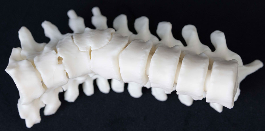
With 3D models, surgeons gain a complete, 360-degree view of the spine before heading into the operating room. Your child's surgeon can flip the model around and study it from every angle. Seeing the spine from front to back and on both sides helps create a safe and accurate surgery plan.
Our orthopedic surgeons also use 3D models to teach families about certain conditions. Examples include spondylolisthesis and hip disorders.
3D models for heart surgery
3D models have transformed care for the most complicated and rare heart conditions. These children often have a one-of-a-kind puzzle for doctors to solve.
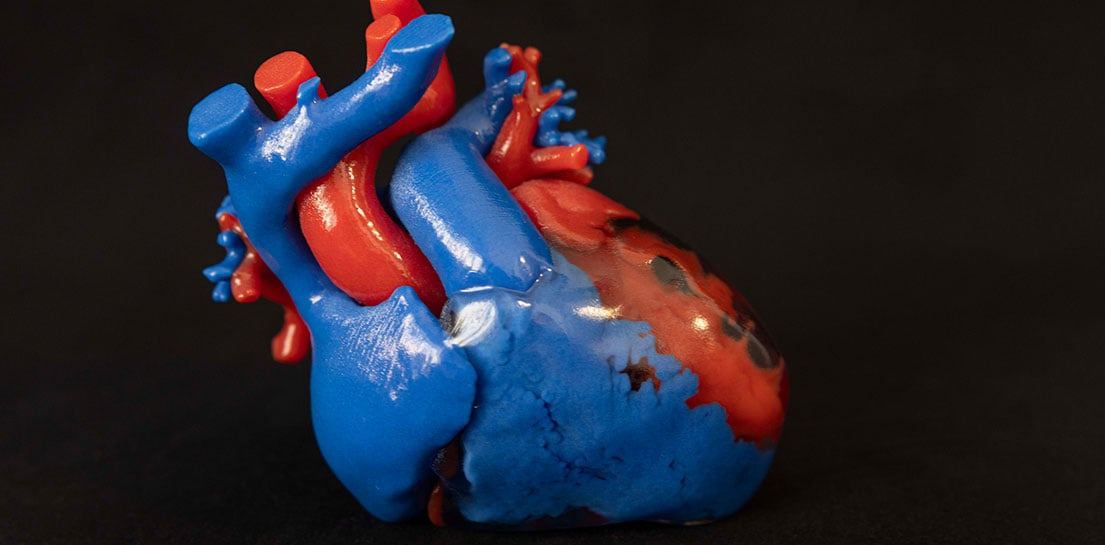
3D models offer surgeons at our Heart Center a window into your child's anatomy. They can rotate models and remove parts to see valves, major blood vessels and other features. Seeing your child's heart in 3D makes it easier to plan for surgery and practice the steps.
Heart conditions we use 3D models for include:
- Complex transposition heart defects, where the blood vessels or pumping chambers of the heart form in the wrong position
- Double outlet right ventricle (DORV), a condition where the aorta connects to the wrong area of the heart
- Heterotaxy heart defects, where the heart and other organs are in the wrong spot in the chest
3D models for craniofacial and other plastic surgery
Medical expertise and creativity often combine during Plastic Surgery at Cook Children's.
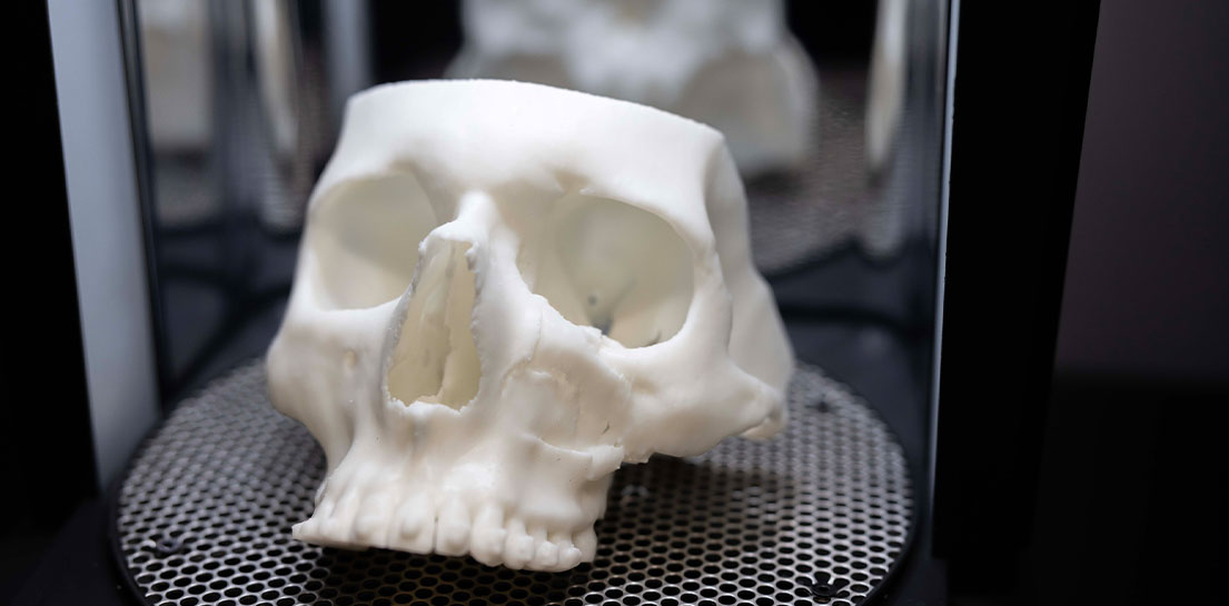
Our surgeons think of innovative solutions, then apply the latest techniques and technology. These powerful tools include 3D printing, for issues caused by:
- Accidents
- Congenital conditions children are born with
- Acquired conditions that develop after birth
Our lab creates models within 48 hours, for timely treatment of trauma. Our surgeons can then work to restore a child's appearance and function.
Some specific ways our plastic surgeons use 3D modeling include:
Facial symmetry surgery
Sometimes only one side of the face gets injured, such as an eye socket fracture. If so, we create a 3D image of the healthy side and then flip it for the model. This method helps surgeons plan repairs and leads to more balanced results.
Skull fractures
3D models act as roadmaps for surgeons during skull repairs. These custom models are particularly helpful for restoring injured areas. Our guides help surgeons place bone or other materials precisely and accurately.
Facial injuries
Car accidents and sports mishaps can injure a child's face. 3D models allow surgeons to study the damage and make a repair plan. Models also reduce the need for time-consuming adjustments during surgery.
Ear reconstruction
Surgeons used to trace 2D pictures on paper to guide ear repairs. Now, technology allows us to scan the unaffected ear, create a 3D mirror image and print models. Surgeons use these models to help craft realistic, matching ears for children.
Cleft lip and cleft palate
3D printing plays a valuable role in some of the treatment for cleft lip and cleft palate. Scans of a child's mouth help us create customized guides. These guides, called splints, support the jaw, palate and teeth in different ways. Surgeons can use them before or after surgery.
Underdeveloped jaws
Surgeons treat underdeveloped jaws (Pierre Robin sequence) by placing a special device to lengthen the jaw. Often, there isn't a lot of room to place this device. With 3D printing, we can craft a replica of a baby's jaw. Our surgeons can then tell exactly where to make incisions and place device hardware while avoiding structures like nerves and tooth buds.
Pediatric surgery and education
There are many other uses for 3D modeling in pediatric surgery. Replicas of the brain help families understand how stroke impacts children, for example.
Recently, our surgeons used 3D models to plan the separation of conjoined twins. Our lab created models of each twin's chest. The surgical team could then practice the incredibly complicated surgery ahead of time.
Get more information about 3D medical models
Call us at 682-885-2140 to schedule an appointment, refer a patient or speak to our staff. We're always happy to answer your questions about our 3D Lab.